 The first of five addresses in the “Deep Space LIVE: Anatomy for All” series of talks by Dr. Franz A. Fellner, head of the Department of Radiology at Linz General Hospital (LGH), was delivered on March 6, 2014 in the Ars Electronica Center Linz. Working together with the Ars Electronica Futurelab enabled Fellner to produce a three-dimensional visualization of the human brain in Deep Space of the Ars Electronica. Further intensive collaboration between Fellner and Futurelab staff researchers is what led up to the series’ second installment, which was presented yesterday: “3-D Patient Data Direct from the Clinic.” We recently chatted with Franz Fellner and Ars Electronica Futurelab Director Horst Hörtner about what we was talking about yesterday, and the enormous potential in medical research of visually depicting the human body’s organs.
 The first installment in the “Deep Space LIVE: Anatomy for All” series at the Ars Electronica Center focused on the human brain. What subject did you treat yesterday?
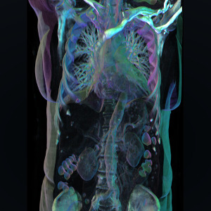
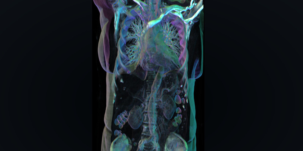
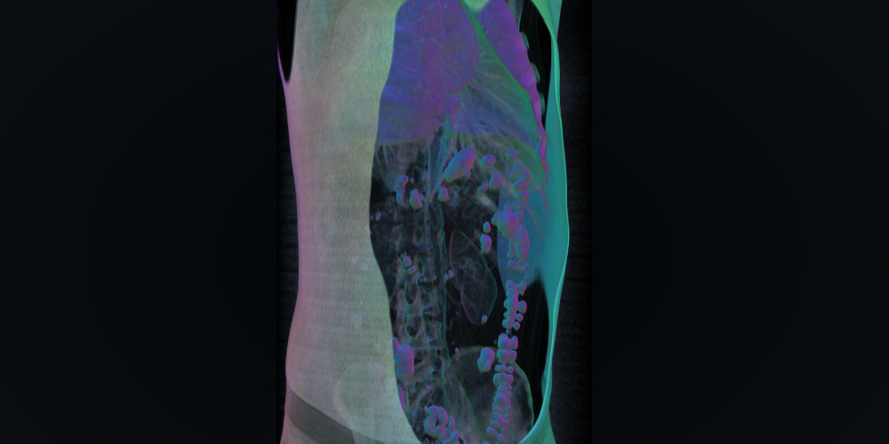
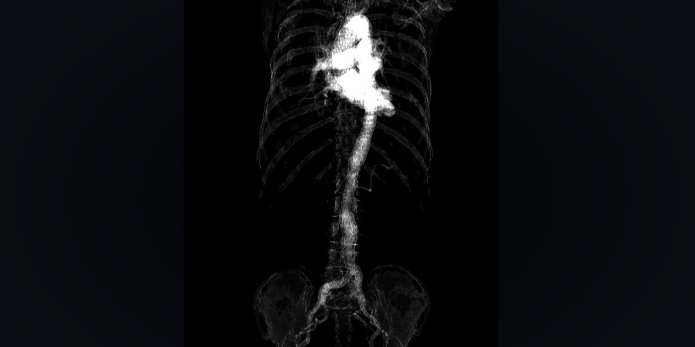
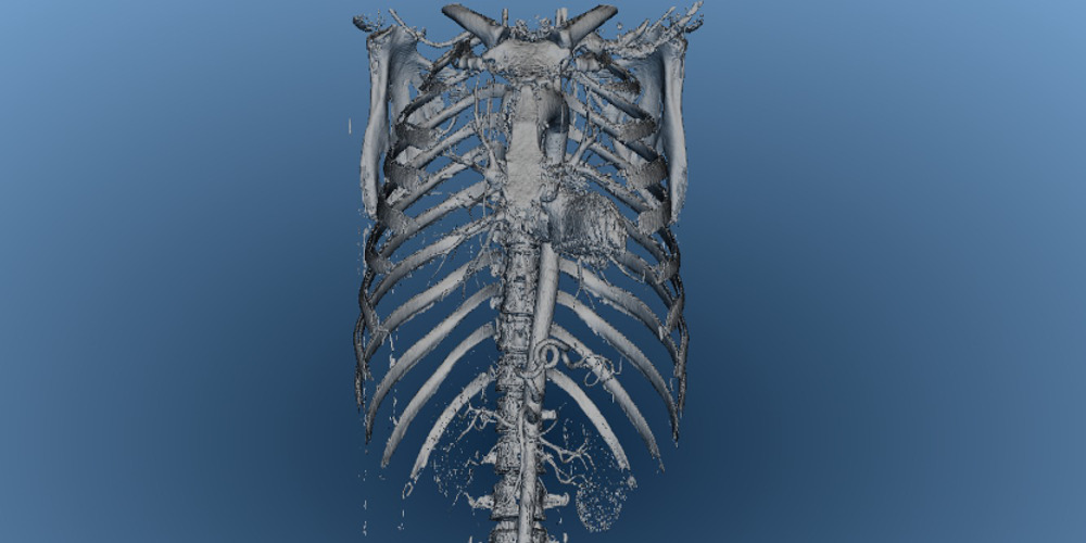
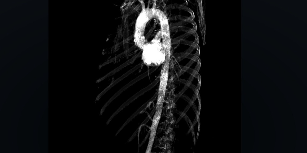
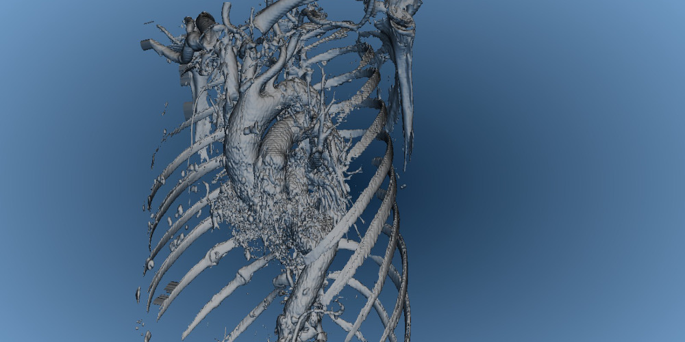
Franz Fellner: Yesterday, we moved down the human body and considered the thorax—that is, the chest and abdomen. We saw which organs are located in this area and how they work. But we were focusing on only a few because there just wasn’t enough time to go into all of them in detail.
 Can you tell us which you were looking at more closely?
Franz Fellner: In the area of the chest, our focus was on the subject of air. How does it enter the human body, where does it go, what does it enable the body to do, and why do we need a steady air supply? In the abdominal area, we dealt with nutrition—beginning with food intake, proceeding on to digestion, and ending up with waste removal.
Last time, you were accompanied by Dr. Michael Malek, a specialist in oral and maxillofacial surgery at LGH, who spoke about future prospects in the field of facial surgery. Did you bring a guest along with you this time too?
Franz Fellner: Yes indeed. And since we had two topics this time—the chest and the abdomen—Iwas supported by two colleagues: Dr. Bernd Lamprecht, director of pulmonology [diseases of the respiratory tract] at LGH, and Dr. Andreas Shamiyeh, a general surgeon and specialist in abdominal surgery at LGH. They showed us what can be done with the visualized radiological data so they can be used directly in conjunction with surgical procedures. This was a highly dynamic and extremely interesting presentation.
And there was a special highlight on tap: live video of a small operation that Dr. Shamiyeh screened at the conclusion of his talk.
 What is the potential of collaboration between LGH and the Ars Electronica Futurelab?
Horst Hörtner: Ars Electronica and LGH are two Linz institutions that have earned worldwide acclaim in their fields; nevertheless, they seem to be working in totally different worlds. But now, we’re in the process of successfully merging these separate areas of great expertise by taking advantage of the extraordinary computer visualization skills available at the Ars Electronica FutureLab and applying them in the field of radiology at LGH. Here, we’re at the forefront in Europe due to the fact that we’re cooperating at a very high level. We now have access to excellent data provided by state-of-the-art radiological equipment, digital material gathered directly from patients that we can input into the visualizations without the need for any sort of data processing. And we can do it not only in a laboratory situation but in front of an audience as well. So this actually is accessible by the general public. And in accordance with Ars Electronica’s tradition of imparting scientific knowledge to laypeople, we’re integrating the expertise of LGH staffers.
What we saw yesterday and what’s been produced here in recent weeks are eye-openers for all of us, to be sure. This has really impressed on us how far this can potentially go.
 What was the particular challenge posed by the visualization of this data?
Horst Hörtner: The challenge in visualizing medical data is that they consist of point data and not of triangulation data as is typical of computer graphics. This is the sticking point that the entire virtual medicine industry is working on today. Computers need triangulated data, or can produce better graphics when they have it. But the results delivered by these imaging techniques are point clouds, and that’s the obstacle. What we’ve now succeeded in doing is to meet this challenge, so to speak. We’ve overcome this hurdle to the extent that it’s unnecessary to introduce an additional process and modify the raw data itself because we’ve discovered a way to concentrate strictly on the visualization. The original data can remain more or less unchanged. We’ve been able to surmount this impediment using the equipment we have here in DeepSpace and the software tools we’ve developed in recent years at the Futurelab. We’ve succeeded in eliminating the need for further data processing and going right to the visualization.
 How can the graphic depiction of the human body’s organs have an impact on medical research? What exactly is the potential here in the field of medicine?
Franz Fellner: The developmental potential is enormous, and, indeed, in two directions. The problem at present is by no means acquiring good imaging; we have that already. Needless to say, it would be great if there were further improvements here, but the real problem is the post-processing. We’re now getting immense qualities of data and we’re viewing them just like we did back in the ‘90s—on tiny monitors. They’ve admittedly gotten a bit larger in recent years, but the process of considering this great wealth of information calls for much more space. It’s a repeated source of fascination to me to realize how much more you can suddenly see by viewing the data on a large-format display instead of on a small monitor. It’s simply because there’s such a wealth of information contained in the data. What we’ve long been dreaming of is to see all of it in 3-D—after all, the things we’re examining are three-dimensional as well. The human body is three-dimensional and we’re constantly visualizing it two-dimensionally. To now be able to see the big picture three-dimensionally on this jumbo-formal display at the Deep Space certainly constitutes a major step forward in the process of disseminating knowledge—to students, medical students and all those who are interested in this field and have to learn about it. This imparts a completely new quality to the process of getting across the true topography of the human body. Perhaps this will also yield applications for medical research. A surgical team will be able to better prepare for difficult operations by having preoperative access to large-format 3-D images. Additional research will have to go into this now.
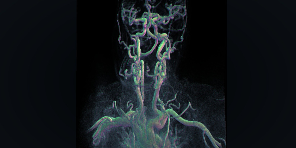 Horst Hörtner: The human being is a visual creature. Our primary means of orientation is our sense of sight. Without a doubt, much has already been accomplished in the processing of the data. When you consider what this technology is already capable of now, it’s simply astounding. Nevertheless, there’s still a lot of potential—for instance, just in the visualizations like we’re doing here in this 3-D space. Maybe, in the future, this will be in an interactive form that will make it possible to manipulate the depicted data and the organs they pertain to; maybe in a way in which you’re no longer seated in front of a screen using a mouse, but rather in a considerably more direct form in which the user can haptically grasp these 3-D visualizations. Eventually, instead of this material being accessible only by the user’s sense of sight, all the senses might get involved. Without being able to predict which illnesses physicians will be better able to treat thereby, I’m nevertheless confident that this will significantly improve the process of diagnosis because physicians will literally be able to grasp illnesses. So what we’re actually doing here is opening a doorway into the realm of comprehending digital information and data visualization in its purest form: 3-D visualization.
Here, there are still a lot of R&D areas that are very closely connected to the various forms of artistic expertise that we’ve been displaying at Ars Electronica over the last 20 years. When we succeed in taking advantage of our skills and know-how, then I see tremendous potential for development here. The artist serves as catalyst at the interface of science and art. Artistically speaking, it’s really a great thrill for me to work with these data. Conversely, there are also significant feedback effects in the field of medicine and on medical technology. And there’s also the high degree of inspiration derived by the scientists involved, who are able to derive new insights from this experience. Thus, this entire endeavor entails a tremendous amount of potential.
Also visit Dr. Franz A. Fellner’s next speeches in the Deep Space LIVE: Anatomy for All series in Deep Space at the Ars Electronica Center Linz and experience, thanks to modern imaging techniques, a fascinating insight into the interior of the human body. For more information visit: https://ars.electronica.art/center/en/programm/anatomie-fur-alle/. Furthermore, on Saturday, June 6. and Sunday, June 29. 2014 the Science Days at the Ars Electronica Center takes place, where you can learn about the human anatomy in detail in an easy new way.