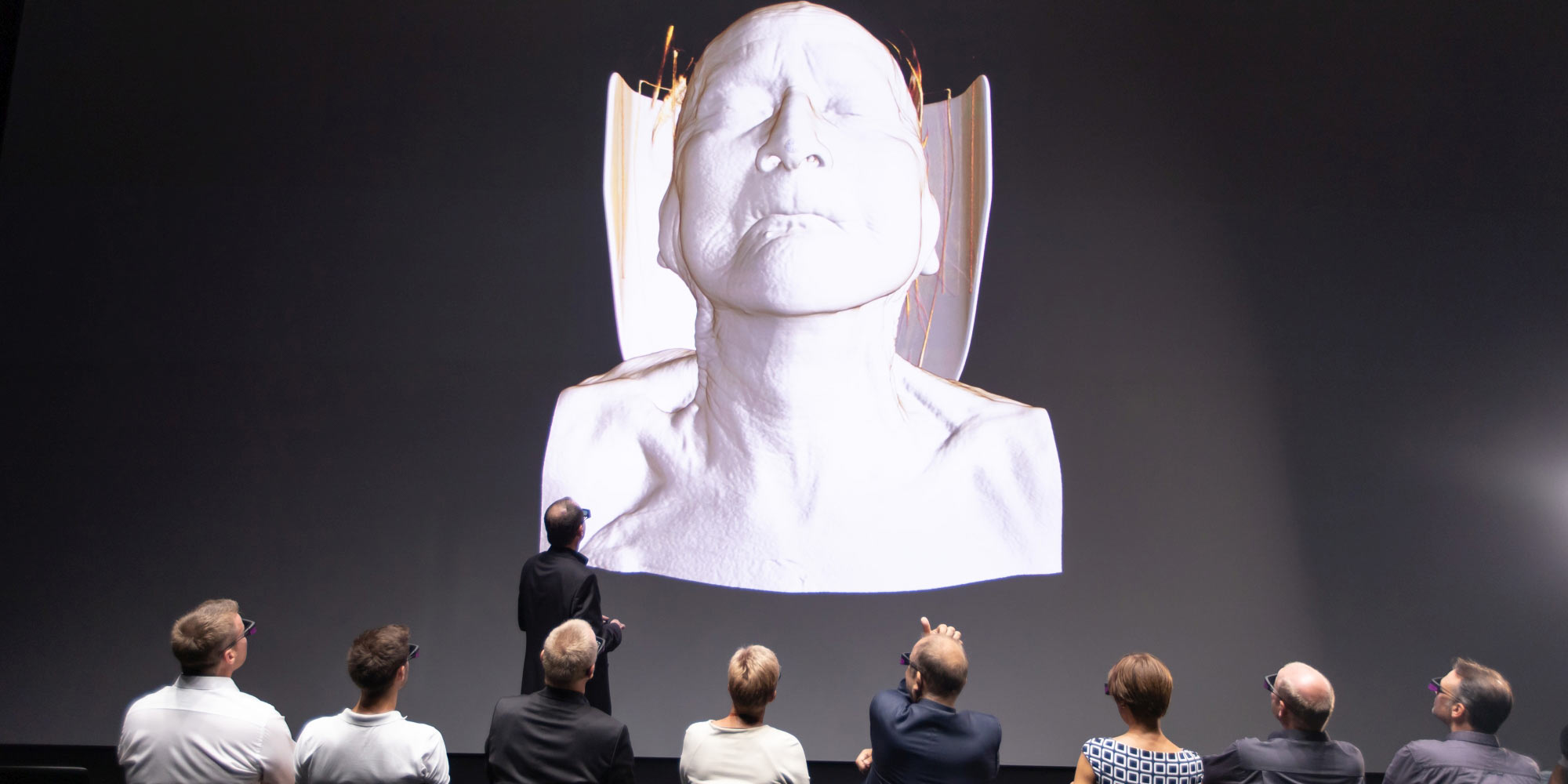
Experience a whole new level of fascination and awe for the human body up close thanks to the software “Virtual Anatomy”, prominently featured in the JKU medSPACE at Johannes Kepler University Hospital in Linz, now open as a venue for teaching anatomy. The Ars Electronica Futurelab is in charge of this prestigious project, one of a kind worldwide, which was created in cooperation with Siemens Healthineers and Dr. Franz Fellner of JKU.
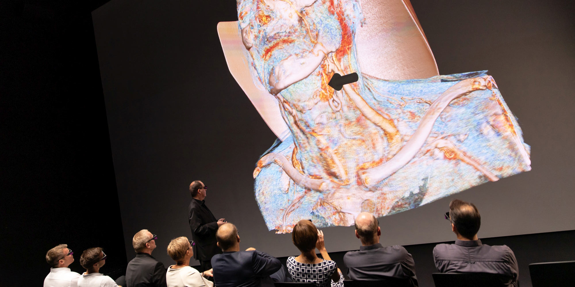
Students can explore the human body in a completely new way in the JKU medSPACE: in high-resolution 3D, zooming and rotating from the outermost layer of skin to the smallest blood vessel — with images that fill the room in 8K resolution on 14 x 7 meters. With “Virtual Anatomy” real radiology data (MRI and CRT images) can be seen as 3D anatomical details of a living human being – instead of abstracted models.
Real data – in real time
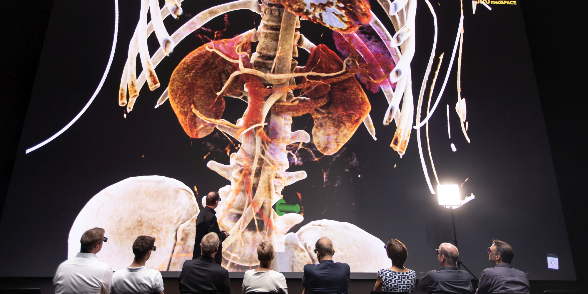
The project is also especially valuable for research and teaching because the Futurelab team has worked for years to develop a way of creating completely new data sets — from brain to whole-body scans — that takes only 15 minutes instead of several hours. This makes it possible for lecturers to independently integrate new data from patients into their teaching, practically in real time.
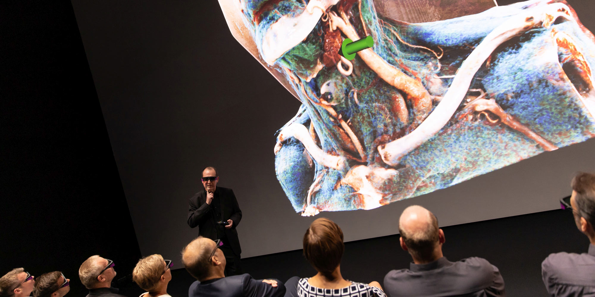
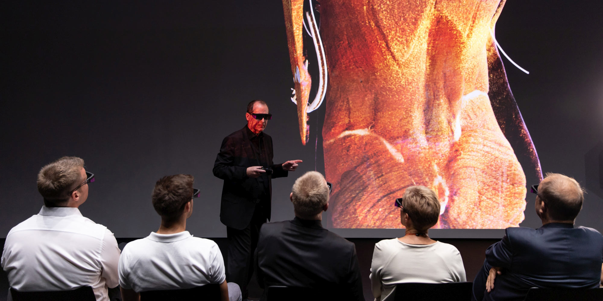
Organs and blood vessels, muscles and tendons, bones and ligaments, as well as tumors and injuries can be magnified to tiniest details as three-dimensional objects in razor-sharp clarity. Students and lecturers can truly immerse themselves in the 3D images and view them from all possible angles. Lecturers use “Virtual Anatomy” to assemble the data they need from computer tomography or magnetic resonance scans and add coloring for illustration purposes.
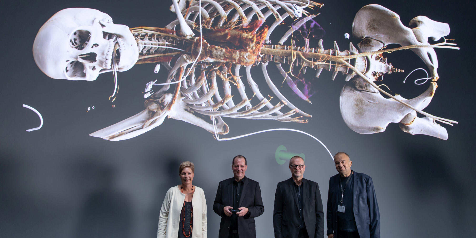
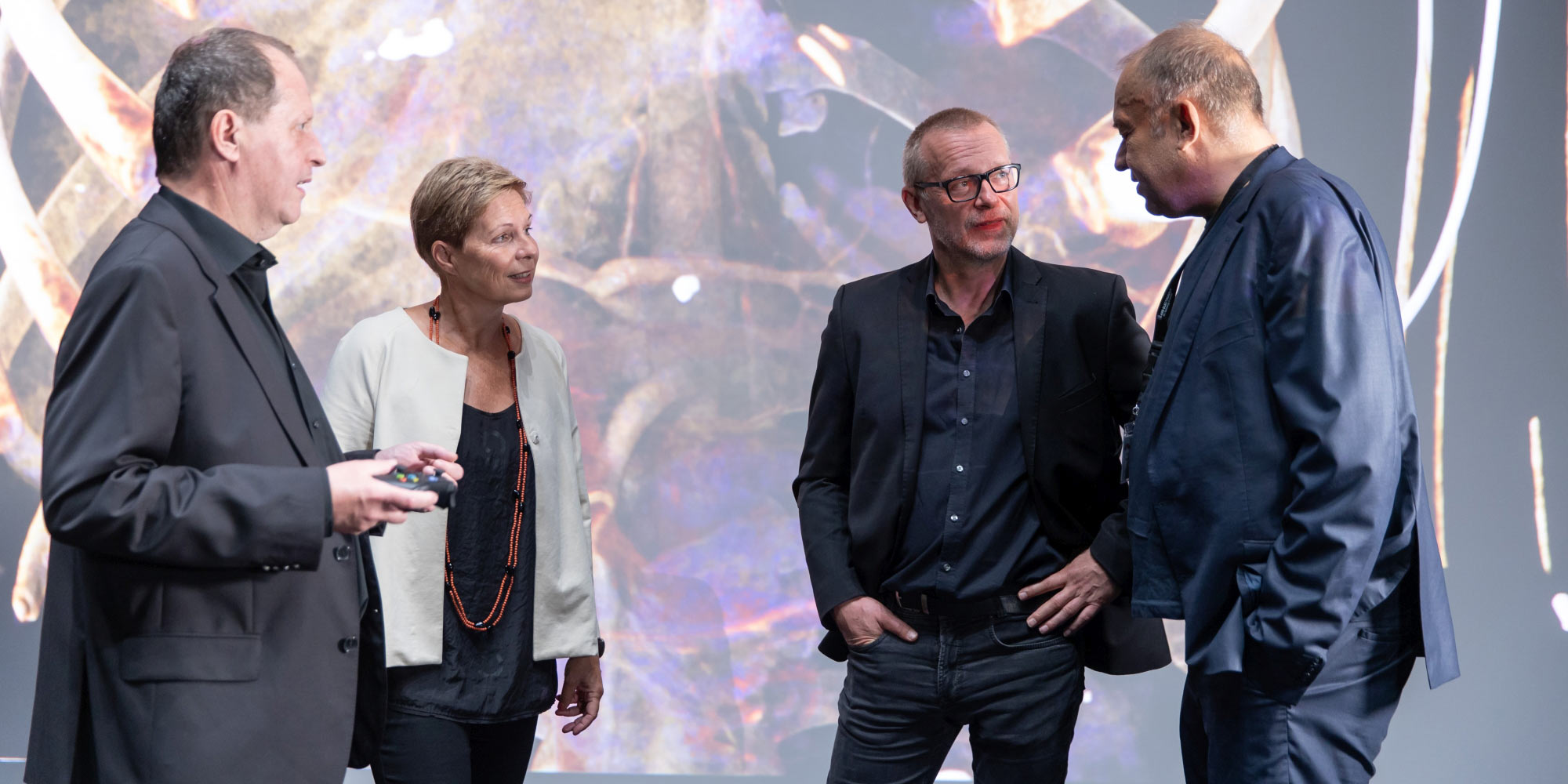
The project was initiated by Dr. Franz Fellner, the current head of the Central Radiology Institute at Johannes Kepler University Hospital, who brought the team led by Siemens researcher Klaus Engel together with the Ars Electronica Futurelab. In September 2015, the 16-by-9-meter Deep Space 8K in the Ars Electronica Center became a lecture hall for virtual anatomy with the software “Cinematic Rendering” for the first time. Regular lectures followed, as did anatomy for amateurs and live feeds of surgeries.
Preparation by transdisciplinary work
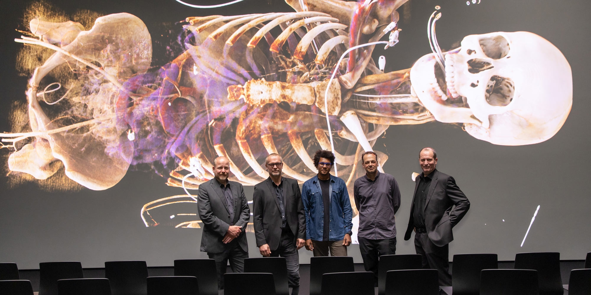
“Virtual Anatomy” and the JKU medSPACE were developed and implemented on the basis of all these experiences. The years-long research project was led by the Ars Electronica Futurelab, with JKU’s medical-didactic expertise building on software from Siemens Healthineers. The lab customized the software to meet the requirements of a modern multimedia lecture hall.
With the JKU medSPACE, the university of the future becomes a reality, preparing medical professionals to operate together with robots, diagnose with AI systems, and teach and learn in virtual environments. The JKU medSPACE also reveals the potential offered by transdisciplinary work of the kind that the Ars Electronica Futurelab has been doing for 25 years — when curiosity and openness, professional and didactic expertise, technological competence and, last but not least, artistic creativity come together.

Want to know more? Read about “Virtual Anatomy” on the Ars Electronica Futurelab website, explore the beginnings of “Cinematic Rendering” on our blog or contact us for more information!