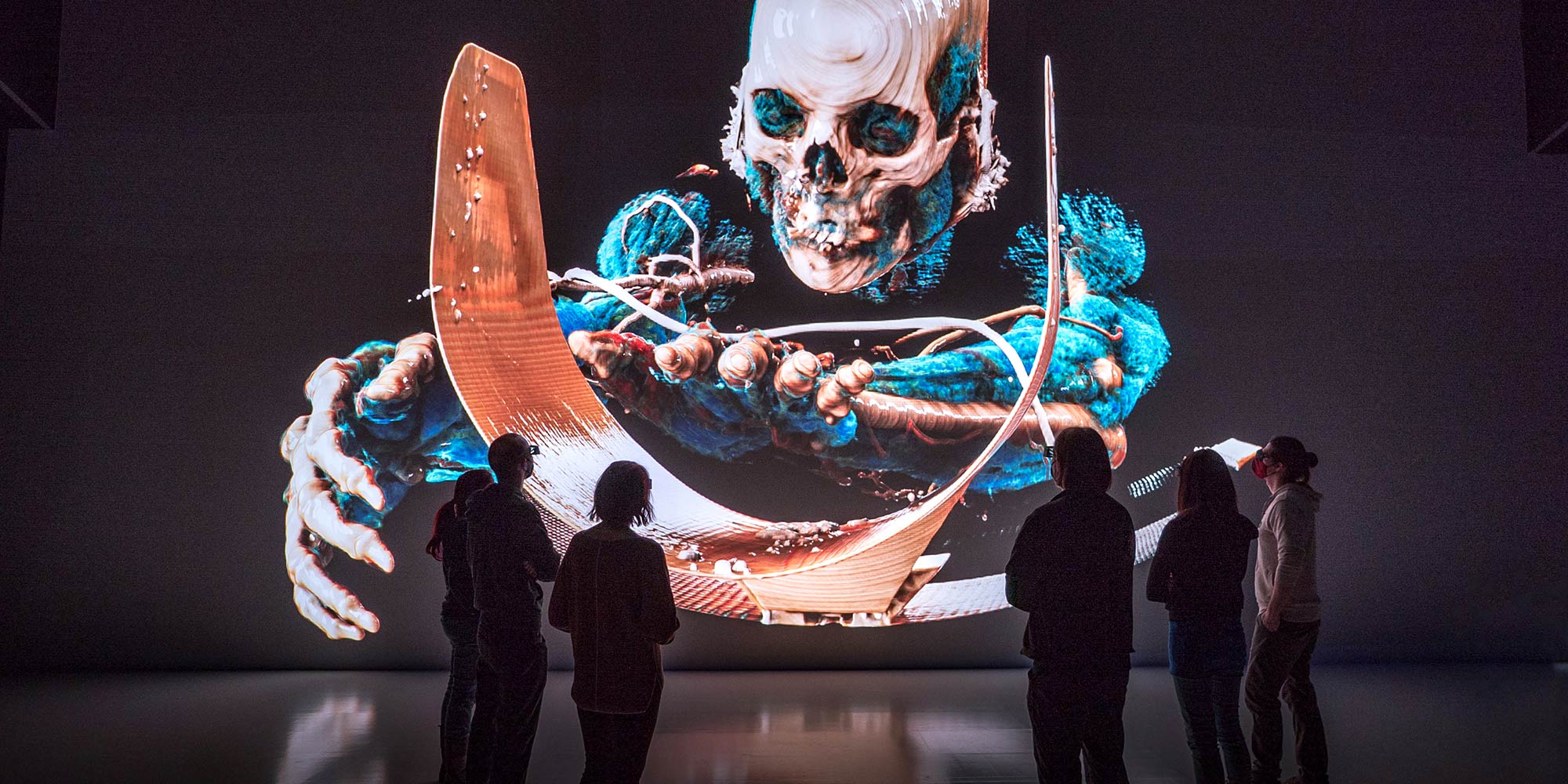
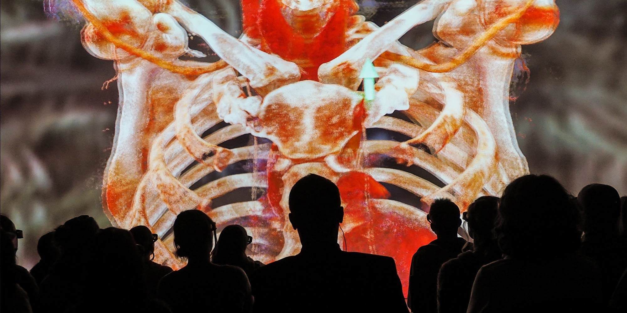
Virtual reality technology meets medical science.
Here, virtual reality technology and medical science come together to display anatomical information stereoscopically and with unprecedented depth of field: Ultra-high resolution projections of anatomical CT and MRI data are displayed in 16×9 metre format.
Virtual Anatomy, photo: Ars Electronica / Martin Hieslmair
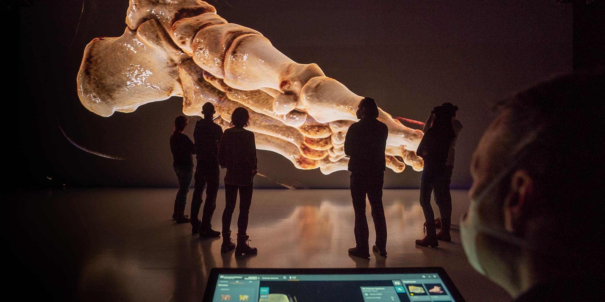
Virtual Anatomy, photo: Ars Electronica / Robert Bauernhansl
Virtual Anatomy was developed in collaboration with Prof. Dr. Franz Fellner from the Department of Radiology at the Kepler University Hospital Linz as Medical Scientific Director. Siemens Healthineers contributed its revolutionary visualisation tool “Cinematic Rendering” to this research project in 2015 in order to use Virtual Anatomy in the education of medical students at Johannes Kepler University Linz.
Finally, in September 2021, the new JKU medSPACE, developed in cooperation with the Ars Electronica Futurelab, went into operation. On the occasion of the opening of JKU medSPACE, the Virtual Anatomy software previously used in the Ars Electronica Center’s Deep Space for anatomy lectures also underwent an upgrade. Now experience stunning insights into the human body, taken to the next level thanks to the new features.