
A look back at the history of medicine quickly reveals that the graphic depiction of the human body and its internal organs has a very long tradition. Beginning with sketches rendered by hand all the way to x-ray techniques and modern digital methods like computer tomography (CT) and magnetic resonance imaging (MRI), the representation of human anatomy and its anomalies has long been an integral component of the field of medicine.
These modern visualization methods provide physicians with powerfully enhanced diagnostic tools, data that constitutes a basis for consulting colleagues about symptoms and prognoses, and thus the ability to prescribe optimal treatments.
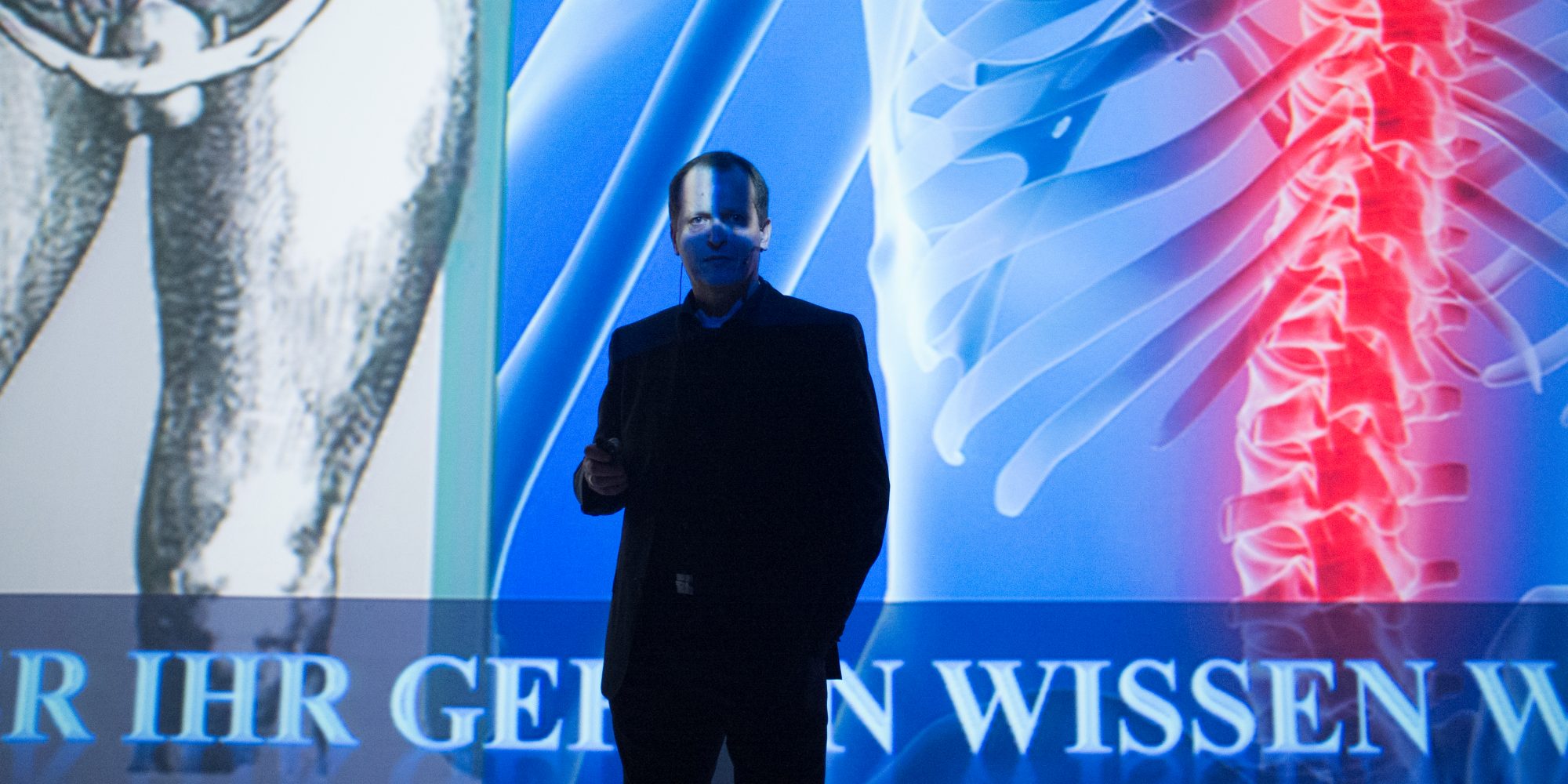
Virtual Anatomy & Pathology Credit: Florian Voggeneder

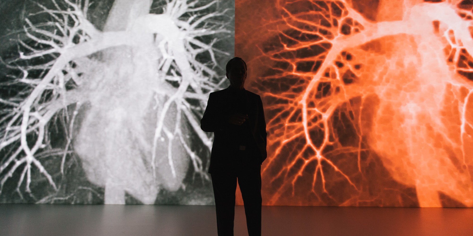
Cinematic Anatomy x Deep Space Credit: Martin Hieslmair
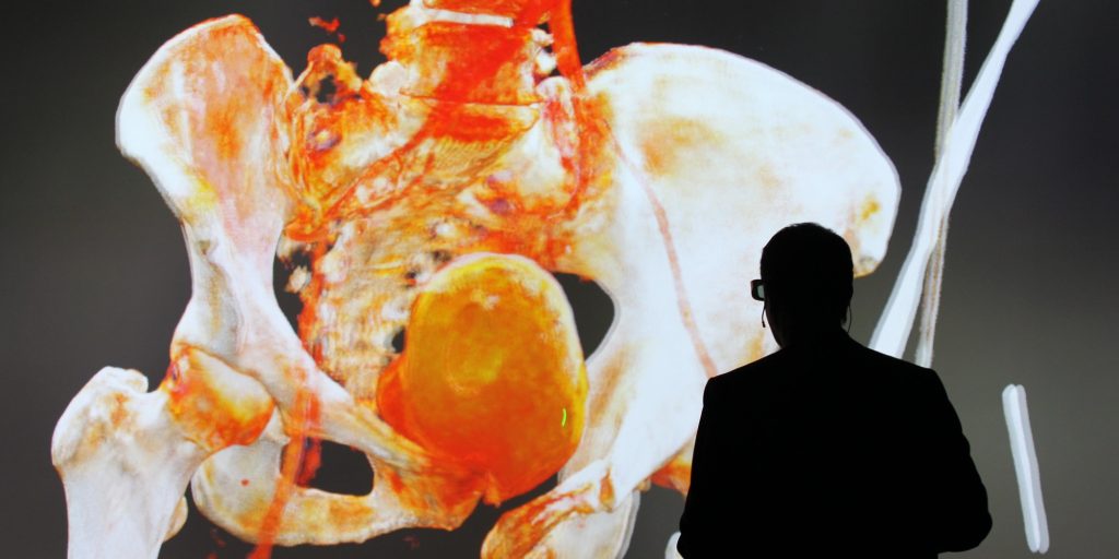
Virtual Anatomy & Pathology Credit: Florian Voggeneder
Univ. – Prof. Dr. Franz Fellner, director of the Radiology Department at Linz General Hospital, and researchers of the Ars Electronica Futurelab have taken the next bold step forward. Their collaborative project “Virtual Anatomy & Pathology (VAP) Deep Space” took available visualization technologies to the next level. The project’s objective was to carry on innovative work being done in the field of digital health by developing new forms of depiction that take innovative approaches to representing the manifold elements of the human body—from entire organs to individual tissue structures. Existing technologies such as ultrasound, CT and MRI already offer a wide variety of superb methods for visualizing the interior of the human body. Accordingly, the challenge taken up by this project was to utilize the data generated by these devices to deliver faster—and, above all, better—visualizations. The attainment of this goal entailed adapting algorithms that were developed to display aesthetically demanding content in the world’s highest-definition projection setting, Deep Space at the Ars Electronica Center.
The results are ultra-high-resolution projections of anatomical CT and MRI data in 16×9-meter format. In addition to their jumbo dimensions and breathtaking sharpness, the images can also be displayed in 3D. This extraordinary combination of virtual reality technology and medical science now makes it possible to depict anatomical information stereoscopically and with a depth of field that has never before been possible in this form using previously available technologies. The advantage of this procedure is not only the ability to capture and depict medical imaging data in much greater detail; this project also imparts powerful new impetus to further medical research on the depiction of human anatomy, physiology and pathology. The next step will be to investigate the extent to which this new technology can also deliver benefits for the planning and simulation of therapeutic measures.
The Universe Within – Cinematic Rendering
Because of the technical innovations in the Deep Space 8K, it is now possible to use highly complex programs. One is the Cinematic Rendering app for the presentation of photorealistic images of the human body in 3-D and jumbo-format dimensions.
Credits
Medical-Scientific Director: Prim. Univ. – Prof. Dr. Franz Fellner (AKh Linz/Department of Radiology)
Software Development and Visualization: Florian Berger